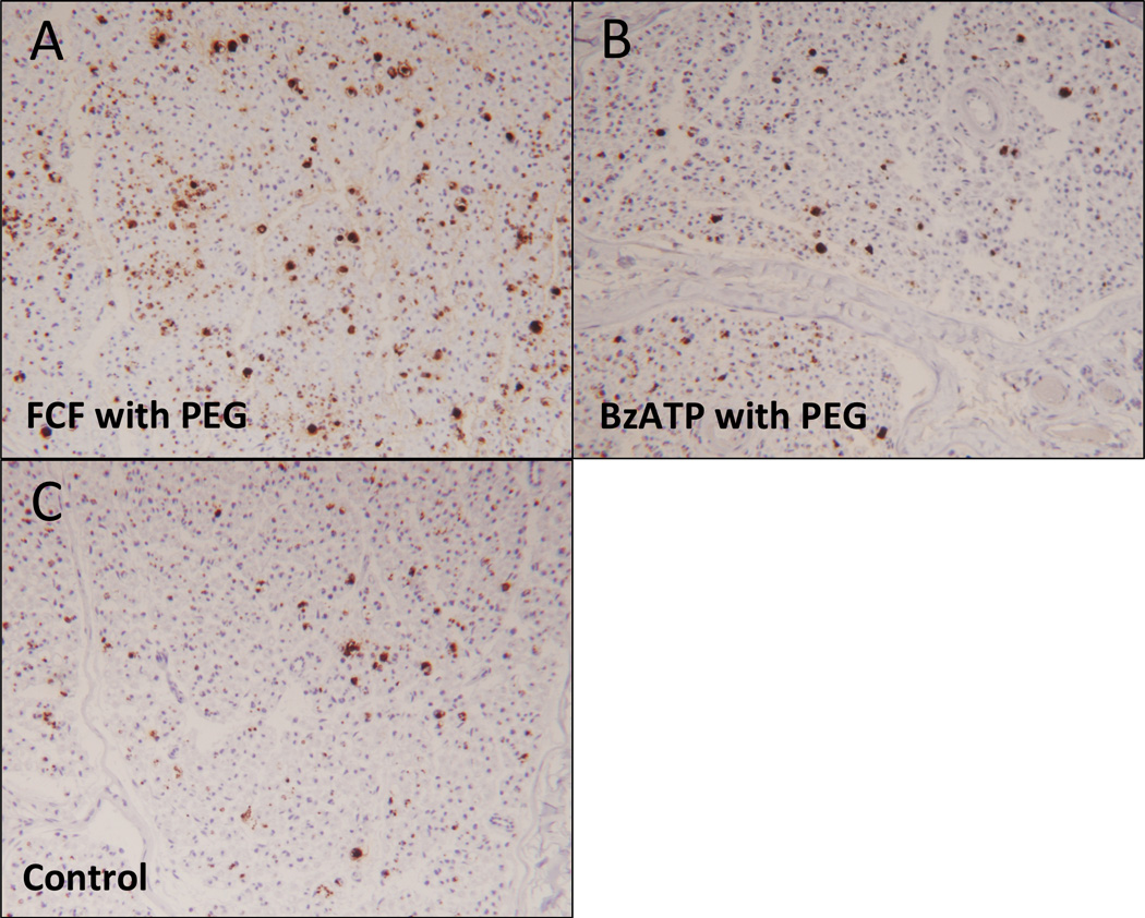
Figure 5.

Representative photomicrographs of myelin basic protein staining, 21 days postoperatively, distal nerve cross section comparing:
A. FCF with PEG treated nerve
B. BzATP with PEG treated nerve
C. Control nerve

Representative photomicrographs of myelin basic protein staining, 21 days postoperatively, distal nerve cross section comparing:
A. FCF with PEG treated nerve
B. BzATP with PEG treated nerve
C. Control nerve