
Figure 6.
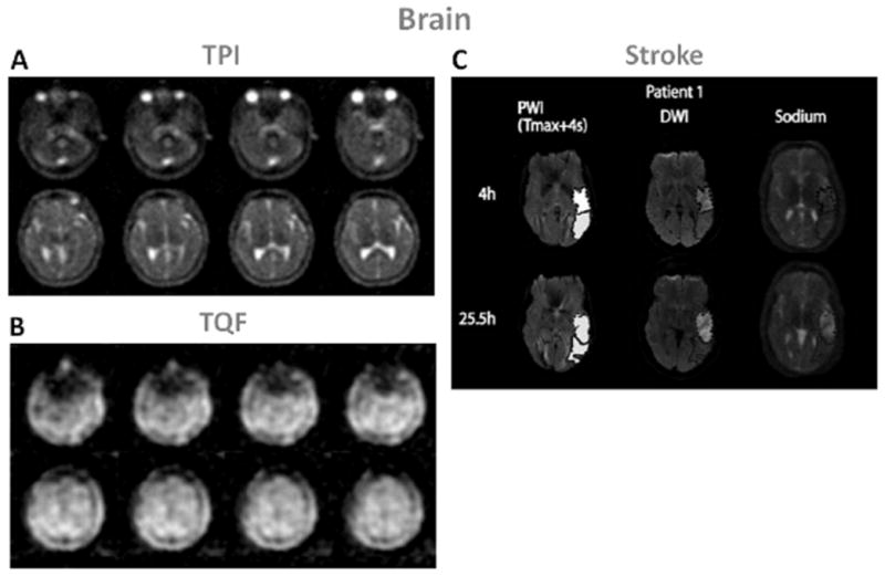
Examples of brain images. A: Twisted projection imaging (TPI) at 3T of a healthy brain. B: Triple-quantum filtered (TQF)-TPI at 3T of a healthy brain. C: Representative perfusion weighted imaging (PWI), diffusion weighted imaging (DWI) at 1.5T and sodium TPI at 4.7T of the brain of a patient with acute ischemic stroke. These images show the hypoperfused (Tmax+4s) perfusion maps, the DWI hyperintense (core) in dotted outline and the PWI-DWI mismatch tissue (penumbra) in solid outline, and corresponding sodium images, acquired 4h and 25.5h after symptom onset. Figures A and B: Courtesy of Professor F. Boada, New York University Medical Center / University of Pittsburgh Medical Center. Figure C from Tsang A. et al. J Magn Reson Imaging 33:41–47, 2011. Reproduced by permission of Wiley-Blackwell.