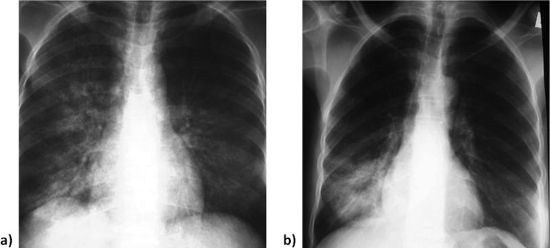
Figure 2.
Radiographic appearance of lung opacities with (a) C. burnetii. Mildly cropped anteroposterior film demonstrates coarse right perihilar and lower lobe linear and reticular heterogeneous opacities. Less well visualized are fine-medium left mid-lung reticular and small nodular opacities. (b) S. pneumoniae. Minimally croppedanteroposterior chest film demonstrates both homogeneous and heterogeneous opacities in the right lower lobe. Centrally the opacity is more uniform or homogeneous, whereas peripherally the pneumonia is more a combination of linear and reticular opacities or heterogeneous. This example was also typical of the type of abnormality seen on chest radiographs in patients with bacterial pneumonia, (e.g., S. pneumoniae).

