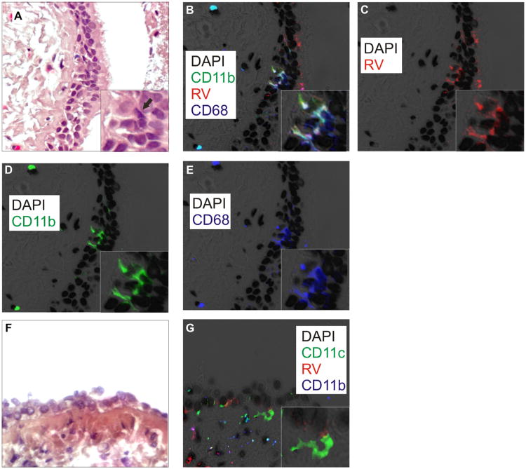
Fig 2.
Asthmatic tissue stained with DAPI (black), R16-7 (red), anti-CD11b (green), and anti-CD68 (blue). A, F, Hematoxylin and eosin. The arrow in A marks a corresponding RV16-, CD11b-, CD68-positive cell. B, Colocalization of CD11b and CD68 is cyan; colocalization of 3 markers is white. C, RV16. D, CD11b. E, CD68. (Original magnification, ×640; insets show group of costained cells.) G, Image showing RV (red), CD11c (green), and CD11b (blue). CD11c-positive stellate cell underlying infected epithelial cell (inset). DAPI, 4′-6-Diamidino-2-phenylindole, dihydrochloride.

