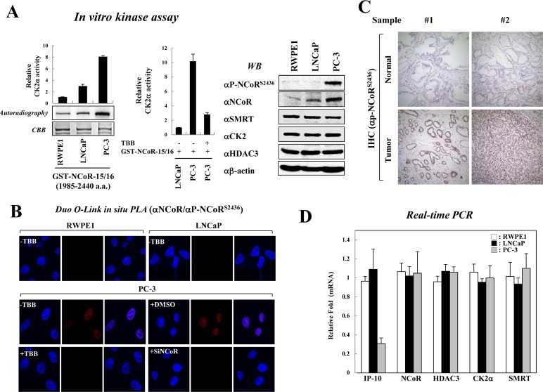
Figure 1. Constitutive activation of CK2 and NCoRS2436 phosphorylation in PC-3 cells.
(A) In vitro kinase assays were performed by incubating GST-NCoR-15/16 protein and immunoprecipitated CK2 enzyme obtained from indicated prostate cancer cells. NCoR phosphorylation levels were analyzed by autoradiography and scintillation counts (left & middle panel). Error bars indicate SD (n=3). Cell lysates from prostate cancer cells were analyzed by Western blotting (right panel). (B) For the Duolink in situ PLA analysis, indicated cells were seeded on coverslips and treated with the siNCoR in the presence of DMSO or TBB (50 μm). The pre-metabolized cells were incubated with the indicated antibodies and treated with PLA probes (PLUS and MINUS). The positive signal was analyzed using confocal microscopy. (C) Expression of NCoRS2436 phosphorylation was examined using immunohistochemical staining in samples from prostate cancer patients. Representative NCoRS2436 phosphorylation levels are shown in two normal and tumor sections. The nuclei were counterstained with hematoxylin. (D) Expression levels of indicated genes from prostate cancer cells were analyzed by real-time PCR. Error bars indicate SD (n=3).

