
Figure 5.
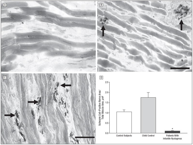
Sections from controls, patients with infantile nystagmus, and a patient with vertical strabismus that have been immunostained for the presence of Schwann cell myelin. A, Typical appearance of muscle from a patient with infantile nystagmus showing extremely sparse innervation (n=10). B, Typical appearance of myelinated nerve fiber content in control extraocular muscles (n=10). C, Typical appearance of myelinated nerves from a patient with vertical strabismus (n=1). Bars are 20 μm. D, Quantification of mean Schwann cell area per total muscle area from controls (n=10), a child control (n=1), and patients with infantile nystagmus (n=10). The 2 muscles obtained from the 7-year-old control are depicted separately to demonstrate their similarity with data from the adult control muscles; however, for statistical analysis, all the control muscles were pooled. *P≤.05. Arrows indicate Schwann cell–positive nerve bundles; error bars, SE.