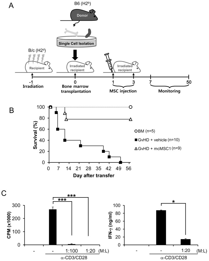
Figure 1.
In vivo efficacy model for MSC. (A) BALB/c mice were irradiated in a dose of 8.5 Gy. Twenty-four hours after irradiation, 5×10⁶ BM and 5×10⁶ spleen cells from B6 female mice were injected. On days 1 and 3 after transplantation, 5×10⁵ MSCs were injected intravenously. (B) Survival rates were monitored. (C) Lymphocytes from the spleen and lymph node were stimulated with anti-CD3 and anti-CD28 antibodies in the presence or absence of mcMSC1, and mcMSC1 was co-cultured with lymphocytes at a ratio of 1:20 or 1:100 (MSCs: lymphocytes). T cell proliferation and IFN-γ production were measured by [3H]-thymidine incorporation and ELISA, respectively. T cell proliferation and IFN-γ assays were repeated three times and similar results were obtained. L, lymphocytes; M, MSCs; BM, bone marrow, ***p<0.005, *p<0.05.

