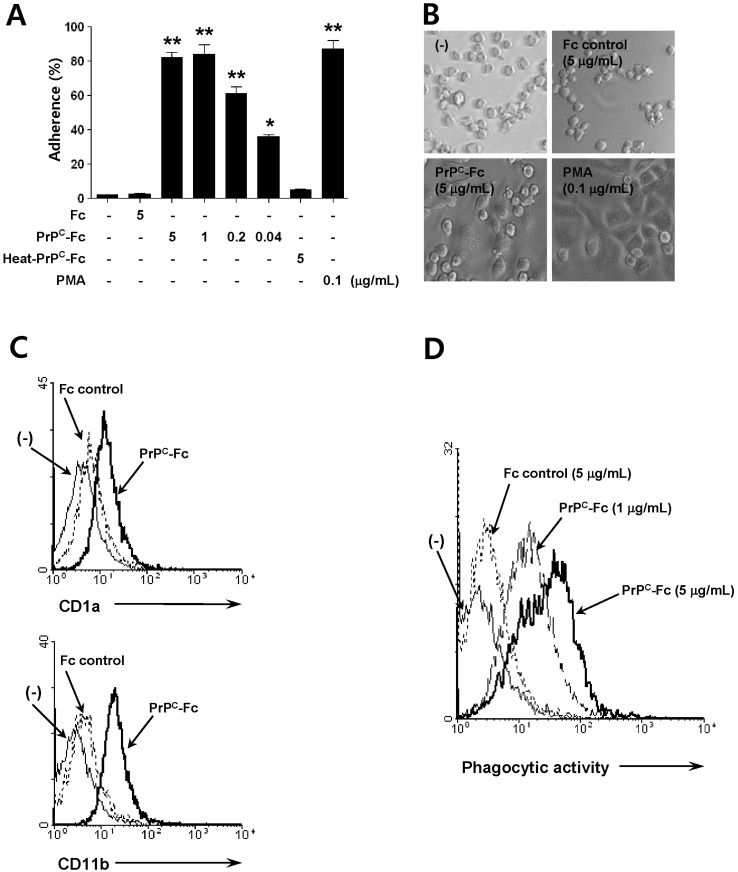
Figure 2.
Differentiation of monocytes into macrophage-like cells following soluble PrPC-Fc treatment. (A) THP-1 cells were treated with soluble PrPC-Fc, heat-denatured (at 95℃ for 30 min) PrPC-Fc, or control Fc at the indicated concentrations for 30 min, and adherent cells were counted. Cell adherence was expressed as a percentage of the total number of cultured cells. The assay was performed in triplicate. Bar graphs represent the mean±SEM. Statistical analysis was performed in comparison with untreated control. *p <0.05; **p<0.01. (B) Phase-contrast images of THP-1 cells treated with soluble PrPC-Fc, control Fc, or PMA for 48 h (200×magnification). (C) Primary monocytes were cultured with soluble PrPC-Fc or control Fc for 24 h followed by labeling with anti-CD1a-FITC or anti-CD11b-FITC and flow cytometric analysis. Similar results were obtained from three independent experiments. (D) Primary monocytes were cultured for 48 h in the presence of soluble PrPC-Fc or control Fc and then incubated for 6 h with E. coli labeled with RFP. Flow cytometric data indicate the amount of phagocytosis of E. coli by monocytes. The data are representative of two independent experiments.

