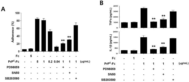
Figure 5.
Role of ERK and NF-κB signaling pathways in monocyte cell adherence and cytokine production induced by soluble PrPC-Fc treatment. (A) THP-1 cells were treated with soluble PrPC-Fc or control Fc at the indicated concentrations for 30 min, and adherent cells were counted. Specific signaling inhibitors such as PD98059 (20µM), SN50 (10µM), or SB203580 (10µM) were added to the THP-1 culture 1 h before soluble PrPC-Fc treatment. Cell adherence is expressed as a percentage of the total number of cultured cells. The assay was performed in triplicate. Bar graphs represent the mean±SEM. Statistical analysis was performed in comparison with soluble PrPC-Fc (1µg/ml)-treated cells. **p<0.01. (B) Primary monocytes were treated with 1µg/ml soluble PrPC-Fc or Fc control for 36 h. Specific signaling inhibitors such as PD98059 (20µM), SN50 (10µM), or SB203580 (10µM) were added to the monocyte culture 1 h before soluble PrPC-Fc treatment. The concentrations of TNF-α and IL-1β in the culture supernatant were determined with the Quantikine Assay Kit. The assay was performed in triplicate. Bar graphs represent the mean±SEM. Statistical analysis was performed in comparison with soluble PrPC-Fc-treated cells. **p<0.01. The data are representative of two independent experiments.

