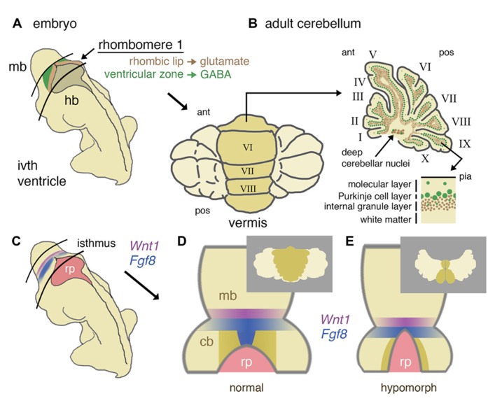
FIGURE 1.
Developmental origins of the mouse cerebellum and the role of isthmic gene expression in patterning the vermis. (A) Schematic representation of a mid-gestation embryo showing the location of derivatives of rhombomere 1. The ventricular layer (green) and rhombic lip (brown) of dorsal rhombomere 1 give rise to all GABA-ergic and glutamatergic cells of the cerebellum, respectively. (B) In a dorsal (posterior) view, the adult cerebellum is characterized by a central (darker shaded) vermis running anterior (ant) to posterior (pos). A uniform layering of cell types can be found throughout the vermis and more lateral hemispheres (shown in schematic parasagittal section), with GABA-ergic and glutamatergic differentially distributed in a later-specific manner: the molecular layer is largely reserved for the interaction of Purkinje cell dendrites and granule cell axons with sparse basket and stellate inhibitory interneurons. The Purkinje cells layer separates the molecular layer from an internal granule cell layer that contains a population of inhibitory Golgi cells. Deep cerebellar nuclei (GABA-ergic and glutamatergic neurons) lie within the white matter. (C) Schematic diagram showing the location of the isthmus organizer at the midbrain/hindbrain boundary with respect to the fourth ventricle roof plate (rp) and the expression domains of Wnt1 (purple) and Fgf8 (blue). (D) Dorsal schematic view of the isthmus region showing with darker shading the approximate region where progenitors of the cerebellar vermis reside, as based on inducible fate-mapping studies (Sgaier et al., 2005). The translation of this dorsal rhombomere 1 territory into adult vermis is shown inset. (E) Altered morphology of the isthmic region and reduced cerebellar size in a hypomorph with an altered function of the isthmic organizer due to diminished FGF signaling. Loss of vermis progenitors is concomitant with the expansion of the roof plate (adapted from Basson et al., 2008). The consequences for vermal morphogenesis in the adult are shown inset.

