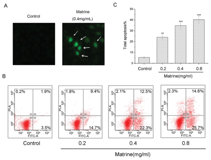
Figure 4.
(A) Morphologically apoptotic changes in M21 cells with Matrine treatment at concentrations as indicated. M21 cells were treated with Matrine for 48 h before TUNEL staining and photographed; (B) Induction of apoptosis in M21 cells with Matrine treatment at concentrations as indicated. M21 cells were treated with Matrine for 48 h before being stained with Annexin V-FITC/PI and flow cytometric analysis; and (C) The total apoptosis in M21 cells with Matrine treatment at concentrations as indicated. All data were expressed as means ± SD of three separate experiments. Significant differences from untreated control were indicated as *p < 0.05; **p < 0.01; ***p < 0.001.

