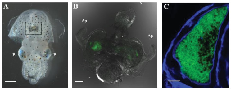
Figure 2.
The light organ of a juvenile E. scolopes harboring V. fischeri. (A) Bright field image showing the ventral side of a juvenile E. scolopes. The dark structure highlighted in the box is the light organ. Scale bar = 1 mm. E = eye; (B) Differential interference contrast (DIC) image of a 48-h p.i. light organ colonized with GFP-labeled V. fischeri cells (green). Scale bar = 100 μm. Ap = appendages; (C) Confocal image of a light organ crypt colonized with GFP-labeled V. fischeri cells (green). Host actin is stained with phalloidin (blue). Scale bar = 10 μm.

