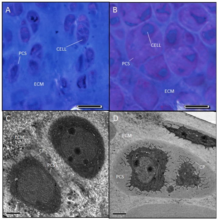
Figure 6.
Two-month-old sedc/sedc mice show detectable metachromasia within the enlarged PCS (B) compared with +/+ mice (A). Electron microscopy revealed a non-fibrillar amorphous material within the enlarged PCS (D). Images were captured at 40× magnification (Bar = 20 μm) for light microscopy (A, B) and 2100× (Bar = 2 μm) for electron microscopy (C,D). Notably, sedc/sedc chrondrocytes show significantly elongated cytoplasmic processes (CP) compared with +/+ mice.

