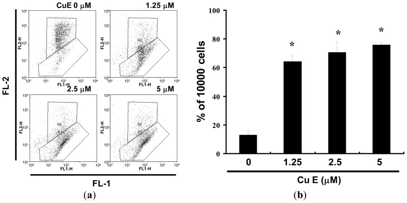
Figure 5.
Reduction of mitochondrial membrane potential (MMP) in SAS cells by CuE, as determined by JC-1 staining and detected by flow cytometry: (a) MMP is shown to be significantly reduced in the SAS cells treated with CuE (0, 1.25, 2.5 and 5 M) by JC-1 staining; and (b) All the data shown are the mean (±SEM) of at least three independent experiments. The symbols (*) in each group of bars indicates that the difference resulting from treatment with CuE is statistically significant at p < 0.05 vs. CuE 0 μM.

