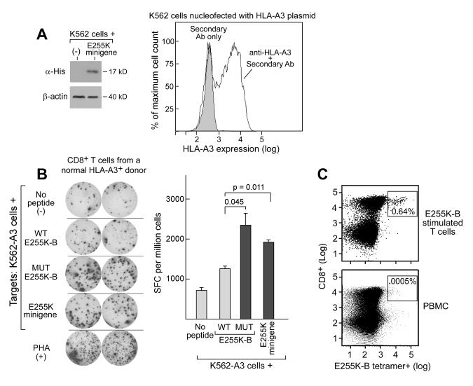
Figure 4. E255K-B is immunogenic in a normal HLA-A3+ volunteer.
A. Western Blot confirming E255K minigene expression (left panel) and flow cytometry confirmation of transient HLA-A3 expression in K562 cells (right panel). B. ELISpot assay results (left panel) demonstrating increased IFNγ secretion following exposure to the mutated peptide E255K-B and the minigene encoding the E255K mutation compared to the parental peptide by a T cell line that was generated following repeated peptide stimulations of PBMC derived from a normal adult volunteer. Shown are duplicate wells for each test condition. APCs used for this assay were HLA-A3+ expressing K562 cells. Right panel - Graphical representation of ELISpot results. SFC-spot forming cells; PHA-phytohemagluttinin. C. Flow cytometric detection of a discrete population of E255K-B-reactive CD8+ T cells within the bulk T cell line by immunostaining with a PE-conjugated E255K-B-HLA-A3+ specific tetramer together with anti-CD8-FITC (top panel), compared to a control population of PBMC (bottom panel).

