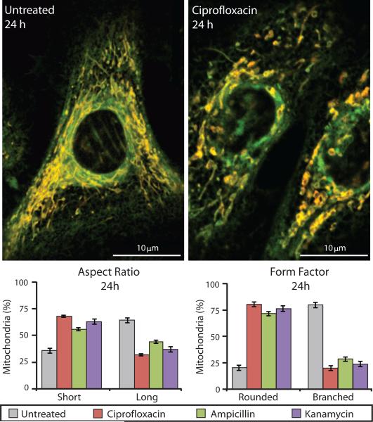
Figure 3. Mitochondria in bactericidal antibiotic–treated cells show an abnormal, profission state.
Mitochondrial morphology was measured in primary human mammary epithelial cells using TMRE and MitoTracker Green. Untreated samples (left image) show mitochondria with normal morphology, which are long and highly branched. Ciprofloxacin-treated cells (10 μg/ml) (right image) had abnormally short and truncated mitochondria. The percentages of mitochondria with a short versus long aspect ratio and rounded versus branched form factor were measured for untreated and bactericidal antibiotic–treated cells. Data are means ± S.E.M. (n ≥ 5).

