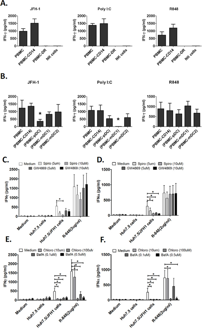
Figure 2. Human NK cells produce IFN-γ in response to HCV-infected hepatoma cells in an accessory cell dependent manner.
(A) Human PBMCs, PBMCs depleted of CD14+ monocytes (PBMC-CD14), PBMCs depleted of HLA-DR+ cells (PBMC-DR) or purified NK cells were co-cultured with JFH-1/Huh7.5 cells or stimulated with poly I:C or R848 for 24 hours, IFN- γ production in the supernatants was measured by ELISA (Mean±SD, n=3). (B) Human PBMCs, PBMCs depleted of CD14+ monocytes (PBMC-CD14), PBMCs depleted of pDCs (PBMC-pDC), PBMCs depleted of mDC1s (PBMC mDC1) or PBMCs depleted of mDC2s (PBMC-mDC2) were co-cultured with JFH-1/Huh7.5cells or stimulated with poly I:C or R848 for 24 hours, IFN-γ production in the supernatants was measured by ELISA (Mean±SD, n=3). *P<0.05 versus other groups. (C, D) PBMCs from healthy donors were co-cultured with JFH-1/Huh7.5 cells or stimulated with R848 for 24 hours in the presence or absence of exosome inhibitors as indicated. IFN-γ and IFN-α production in the supernatants was measured by ELISA (Mean±SD, n=6, *P<0.05). (E, F) IFN-γ and IFN-α release was measured by ELISA in the presence of endosome inhibitors, chloroquine and BafilomycinA. (Mean±SD, n=6, *P<0.05).

