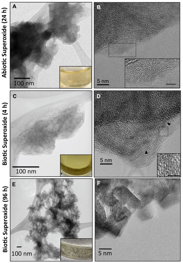
FIGURE 4.
Transmission electron microscopy (TEM) images (A,C) illustrating small dispersed, colloidal Mn oxides formed via oxidation of Mn(II) by abiotically (A) and biotically generated (C) superoxide and 24 and 4 h, respectively. TEM images of Mn oxides formed by biotic superoxide after 96 h show mineral particle aggregates (E). Texture underneath the particles is due to the lacey-carbon support film. Insets in (A,C,E) are pictures of the reacted solution appearance. Biotic superoxide Mn oxides were formed via reaction of superoxide-generating microbial cell-free filtrate with Mn(II) as described in detail previously (images modified from Learman et al., 2011b). High resolution TEM (B,D,F) demonstrating poor crystallinity of the superoxide-produced Mn oxides with a lack of discernable lattice fringes in abiotic generated Mn oxides (B) and small (~5 nm) crystalline domains in biotic Mn oxides (D). HR-TEM images of Mn oxides formed by biotic superoxide after 96 h, however, show defined crystalline domains (F). Insets in (B,D) illustrate the difference in crystallinity (scale bar = 2 nm).

