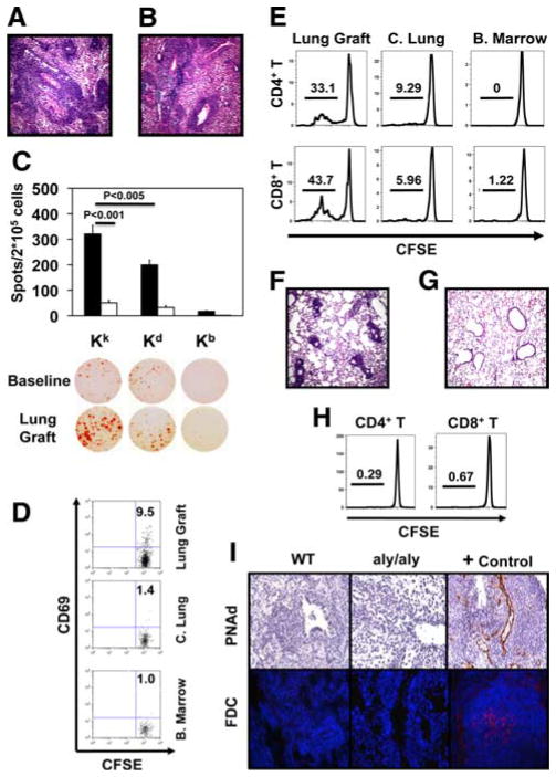
FIGURE 3.
Lung transplantation in the absence of secondary lymphoid organs. A, C3H → splenectomized B6 aly/aly lung grafts on postoperative day 7. B, B6C3F2 Kk/b aly/aly → splenectomized B6 aly/aly lung grafts on postoperative day 7. C, Frequencies of CD8+ T cells isolated from spleens of untrans-planted B6 aly/aly mice (n = 3) or from B6C3F2 Kk/b aly/aly → splenectomized B6 aly/aly lung transplants (postoperative day 7) (n = 5) producing IFN-γ after culture with donor-type (Kk), recipient-type (Kb), or third party (Kd) stimulators. D and E, CD69 expression on T cells in lung grafts (11.2 ± 0.7%) vs contralateral lungs (C. Lung; 2.5 ± 0.6%, p < 0.05) and bone marrow (B. marrow; 2.8 ± 0.9%, p < 0.05) at 30 h (n = 4) and T cell division 3 days after injection into B6C3F2 Kk/b aly/aly → splenectomized B6 aly/aly transplants. F, B6C3F2 Kk/b aly/aly → splenectomized B6 aly/aly lungs on postoperative day 3. G, B6 aly/aly → splenectomized B6 aly/aly lungs on postoperative day 7. H, Proliferation of adoptively transferred T cells in lung grafts 3 days after B6 aly/aly → splenectomized B6 aly/aly transplantation. Data are representative of at least three independent experiments. I, Immunostaining for PNAd (top; original magnification ×200) and FDC (bottom; original magnification ×400) on C3H → B6 and B6C3F2 Kk/b aly/aly → splenectomized B6 aly/aly lung grafts on postoperative day 7. Positive and isotype control Ab staining shown on axillary lymph node (PNAd) and spleen (FDC) from wild-type (WT) B6 mouse.

