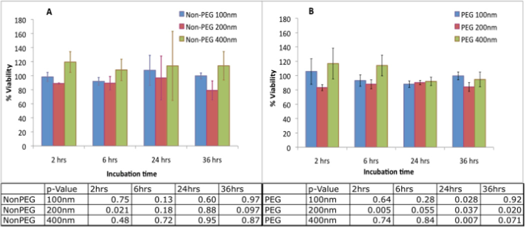
Figure 3.
Effect of liposomal-iodine uptake on cell viability of RAW 264.7 macrophages. The macrophages were incubated with non-PEG liposomal-iodine (A) and PEGylated liposomal-iodine (B) for different incubation times and the cell viability assessed using the MTS assay. Error bars represent standard deviation. Untreated cells were used as controls and their viability numbers were averaged and normalized to 100%, and all other test cases normalized against this standard.

