
Figure 3.
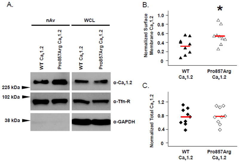
Increased Surface Membrane Density of Cav1.2 Pro857Arg Channels. HEK293 cells expressing either YFP-tagged Cav1.2 WT or Cav1.2-Pro857Arg along with auxiliary β2cN4 and α2δ1 subunits were cell surface biotinylated at 4°C. (A) NeutrAvidin (nAv)-captured surface membrane proteins (left panels) and whole cell lysates (WCL, right panels) were analyzed by SDS-PAGE and western blotting. Densitometric analysis of surface membrane Cav1.2 signal (B) or total cellular Cav1.2 (C) was performed to summarize the results of nine independent experiments. In both cases, normalization of Cav1.2 signal was achieved using the corresponding signal of the endogenous surface membrane protein transferrin receptor (Tfn-R). Solid red lines through distributions indicate population means. Data were compared using Student’s unpaired t-test and * denotes p ≤ 0.05 when WT-Cav1.2 was compared with Pro857Arg-Cav1.2.