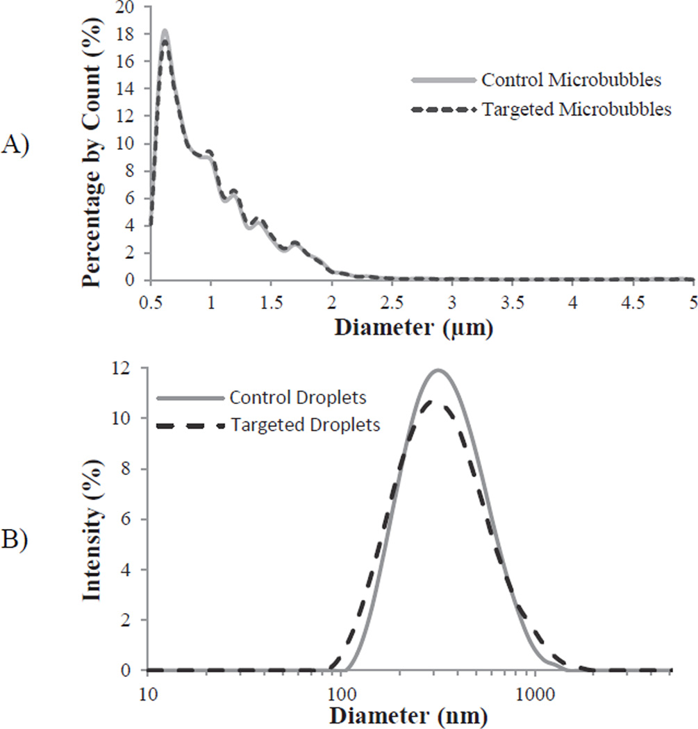
Figure 3.
Representative sizing of A) precursor microbubbles and B) targeted and non-targeted droplets. Note: microbubbles were measured with laser-light diffraction (Accusizer) and are presented as count-weighted distributions, while droplets are measured with dynamic light scattering (ZetaSizer) and are presented as intensity-weighted distributions.

