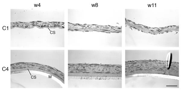
Fig. 1.
Light micrographs of 2 representative constructs (C1 and C4) over time. Cross-sections are shown after 4, 8, and 11 wks of culture. C1 is shown without the transwell membrane, which was removed before processing for evaluation. C4 has the transwell membrane (M) in place. CS, collagen substrate deposited on the membrane prior to seeding the cells. Scale bar = 50 μm.

