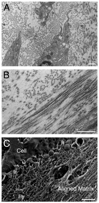
Fig. 2.
Transmission electron micrographs of construct (C4) after 8 weeks. A: Representative image of collagen fibrils and matrix surrounding cells. Note the presence of RER in cells. B: Representative image of lamellar-like architecture and alternating arrays in the construct. C: Representative electron micrograph of cell and the extracellular matrix prepared by Quick Freeze Deep Etch (QFDE). Note that an aligned matrix is present. Scale bar = 500 nm.

