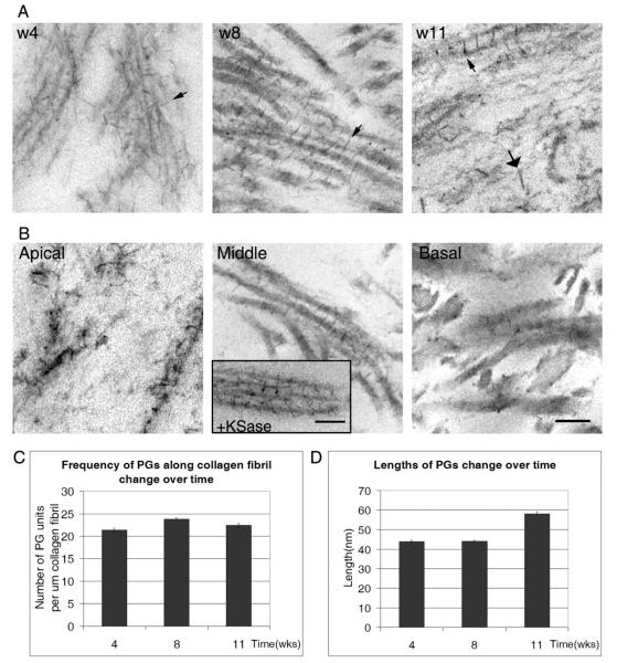
Fig. 7.
Transmission electron micrographs of Cuprolinic blue dye stained proteoglycans in constructs (A, B). A: Four, 8 and 11 weeks. Small arrows show axial alignment of filaments (w4 and 8) and large arrows indicate large filaments (w11). B: Electron micrographs at 8 weeks of construct 4 throughout the depth of the construct. Inset: Electron micrograph showing the presence of filaments after digestion with keratanase and the appearance of short filaments. C: Calculation of PGs along the collagen fibril. D: Change in length of PGs over time. Scale bar = 100 nm.

