FIG 2 .
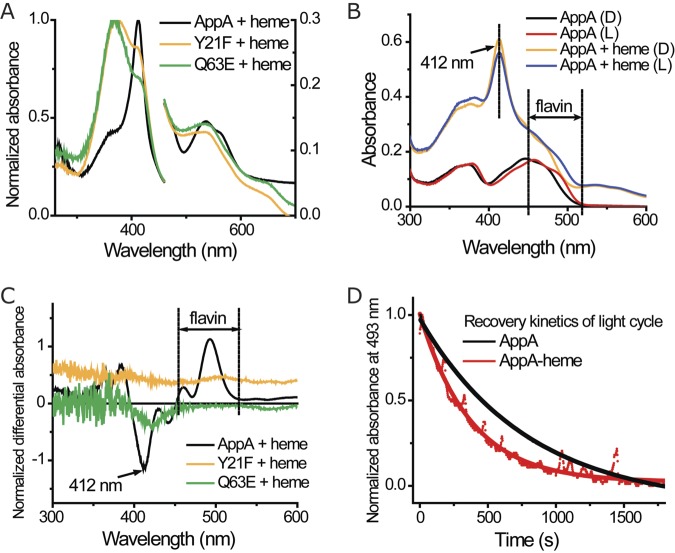
Integration of light sensing and heme binding in AppA. (A) Absorption spectrum of heme and different AppA constructs under aerobic conditions. AppA mutants and heme were mixed at a 2:1 ratio and incubated for at least 20 min before the spectrum was taken. The spectrum of the protein with flavin is subtracted. Normalized absorbance is shown on the right and left y axes. (B) Absorption spectrum of AppA and AppA-heme complex in dark (D) and lit (L) states. The spectrum of protein was not subtracted. (C) Differential spectrum of AppA mutants and heme (dark state minus light state). The samples in Fig. 2A were used here. (D) Recovery kinetics of AppA/AppA-heme light cycle. AppA and AppA-heme complex were light excited and put back in dark conditions. Absorbance at 493 nm was measured to monitor the photocycle of the BLUF domain, and the data were fitted to a single-exponential model.

