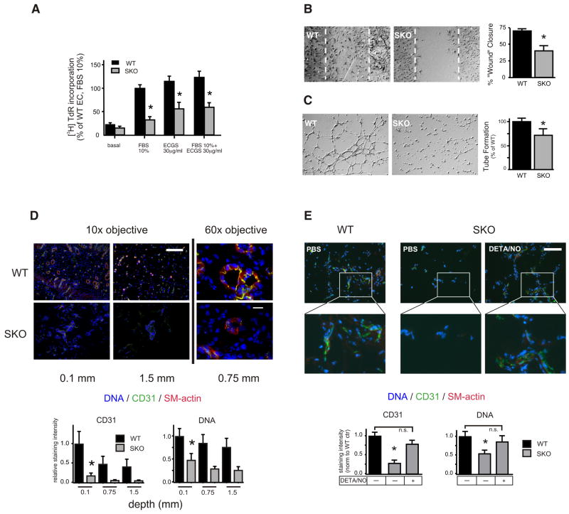
Figure 4. Blunted proangiogenic properties of SKO ECs in vitro and defective capillary formation in vivo.
Primary murine ECs were isolated from WT and SKO mice and were assayed for: (A) [3H] thymidine incorporation in response to different concentrations of FBS with or without endothelial cell growth supplement (ECGS). (B) The chemokinetic response to 1%FBS using an in vitro wound repair (Scratch-) assay (24h), (C) EC tube formation on Matrigel matrix. All experiments were repeated at least three times with different primary isolates and performed in duplicate. Representative images are shown in (B) and (C). The stippled area in (B) denotes the extent of the originally denuded area. *p<0.05 vs WT. (D) Matrigel plugs were implanted into WT and SKO mice (n=3) and harvested after 16 days. Recovered plugs (two for each animal) were cryoembedded and stained for the EC marker CD31 and smooth muscle actin (SMA). Nuclear DNA was visualized with Hoechst 33342 (10x magnification, scale bar=200μm, *p<0.05 WT vs SKO, n=6 plugs each genotype). Representative images (60x magnification, scale bar=20μm) are shown, CD31 (green), SMA (red), DNA (blue). (E) Matrigel plugs were implanted into WT or SKO mice. Mice received DETA/NO (2 mg/kg, i.p.) or saline three times weekly for two weeks. Plugs were harvested 16 days post implantation, cryoembedded and stained as in (D) at a depth of 1 mm. Scale bar=200μm. *p<0.05 vs SKO +DETA/NO.

