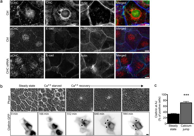
Figure 1.
AJ formation induces CHC phosphorylation and the recruitment of clathrin at cell-cell contacts a. Jeg3 cells were left untreated (top), incubated with non-targeting (middle) or CHC-targeting (bottom) siRNA sequences, seeded and allowed to form AJs for 12 to 16 hours. Cells were then fixed and labeled for immunofluorescence as indicated. b. Representative images (acquired live) of MDCK cells transfected with CLC-GFP and subjected to the calcium jump assay to follow clathrin recruitment at cell-cell contacts (arrowheads) during the formation of adherens junctions. c. MDCK cells at steady state or 12h after the calcium jump were fixed and labeled for clathrin, E-cadherin and actin. The frequency of clathrin recruitment at cell-cell junctions was assayed by fluorescence microscopy. In c values are means (± standard deviation) of three independent experiments. Asterisks represent p values (***= p ≤ 0.001, Student t-test). Scale bars 10 μm.

