Figure 6.
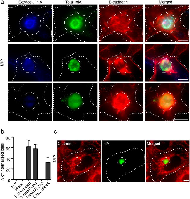
Cell adhesion mediates cell-in-cell events. a. Maximum intensity projections (MIP) of image stacks acquired along the z-axis of HeLa cells transfected with the InlA/E-cadherin chimera (long-dashed outline), trypsinized and incubated for 1 h on a confluent layer of Jeg3 cells (short-dashed outline). HeLa cells were labeled for InlA before permeabilization (blue) to detect extracellular cells and after permeabilization (green) to detect total HeLa cells. Jeg3 cells were labeled for E-cadherin (red). Images represent different stages of cell-cell internalization. b. HeLa cells were either left untransfected (N.T.), transfected with an empty vector (mock), with the InlA/E-cadherin chimera (InlA/hE-cad) or with GFP-tagged E-cadherin (E-cad/E-cad). Cells were trypsinized and incubated for 1h on adherent Jeg3 cells. As a control, HeLa cells transfected with the InlA/E-cadherin chimera were trypsinized and incubated for 1h on ELB1 cells expressing mouse E-cadherin (InlA/mEcad). To test the role of clathrin in cell-in-cell events HeLa cells transfected with the InlA/E-cadherin chimera were incubated for 1h with adherent Jeg3 cells where clathrin heavy chain (CHC) had been previously knocked down by siRNA. In all cases the frequency of cell-in-cell events was quantified by fluorescence microscopy were HeLa cells were differentially labeled before and after permeabilization. Values are means (± standard deviation) of three independent experiments where approximately 100 cells were analyzed for each condition. c. Maximum intensity projections (MIP) of image stacks acquired along the z-axis of HeLa cells transfected with the InlA/E-cadherin chimera (long-dashed outline), trypsinized and incubated for 1 h on a confluent layer of Jeg3 cells (short-dashed outline). Cells were labeled for InlA (green) and clathrin (red). Scale bars 10 μm.

