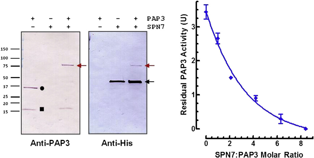
Fig 6.
Formation of serpin-protease complex and inhibition of PAP3 activity by serpin-7. Left: Purified active PAP3 was incubated with serpin-7 (~1:3 protease:serpin molar ratio) for 15 min. Samples were subjected to SDS-PAGE and immunoblotting. Proteins were visualized by using antibodies against PAP3 or hexahistidine tag of the recombinant serpin-7. Symbols represent: PAP3-Serpin-7 complex, red arrow; non-complexed intact form of serpin-7, black arrow; catalytic domain of PAP3, circle; clip domain of PAP3, square. Right: The reaction mixtures were incubated at room temperature for 15 min before adding IEAR-pNA as a substrate. The residual PAP3 activity was measured as the rate of increase in absorbance at 405 nm (Mean ± SD, n = 3). (For interpretation of the references to colour in this figure legend, the reader is referred to the web version of this article.)

