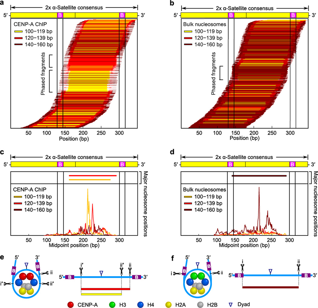
Figure 5. Terminally unwrapped CENP-A nucleosomes and their conventional counterparts with wrapped termini are similarly phased at normal centromeres.
(a,b) The position of each individual CENP-A (a) or bulk nucleosome (b) along a dimerized α-satellite consensus sequence is indicated by a horizontal line. Each fragment is color-coded based on length, as indicated. (c,d) The midpoint positions of CENP-A (c) or bulk nucleosome (d) fragments along the dimer α-satellite consensus sequence. Solid vertical lines indicate the location of the 17 bp CENP-B box (B) in (a–d). (e,f) Models of the preferred positioning and MNase cleavage sites on CENP-A (e) and bulk (f) nucleosomes at normal centromeres.

