
Figure 1.
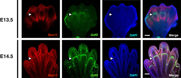
Expression patterns of Sox11 and Gdf5 during joint formation in mouse embryos. Double fluorescence section section in situ hybridization in hind limbs of mouse embryos (E13.5, 14.5). Confocal microscope images show (from left to right): Sox11 (red channel), GDF5 (green channel), DAPI staining of nuclei, and merged images. White arrowheads indicate the location of the interzone at prospective joint sites. Scale bars, 200 μm.