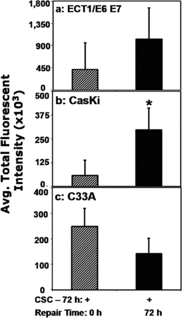
Figure 3.
Assessment of p53 expression in different human cervical cells. ECT1/ E6 E7 (a), CaSki (b), and C33A (c) cells were treated with CSC (12 µg/ ml) for 72 h to induce the 8-oxodG lesion. Then, cells were freed of the residual CSC and further incubated for 72 h for repair of oxidative DNA damage to occur, stained with Alexa Flour 488-conjugated anti-p53 antibody and quantified by flow cytometry.

