
Figure 4. Absence of PD-L1 results in higher activated CD4+ T cells in CNS of E2-implanted mice.
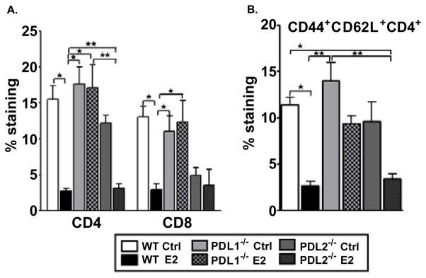
Mononuclear cells isolated from brains of control and E2-implanted WT, PDL1−/− and PD-L2−/− mice were analyzed on Day 24 post-immunization. A) Frequencies of CD4+ and CD8+ T cells were determined in individual brains and indicate the percentages of total gated live leukocytes (n=3–4). Data are pooled from 2 independent experiments (mean ± SEM). B) Flow antibodies for CD44 and CD62L were used to differentiate between naïve, memory and activated (CD44+CD62L+) CD4+ T cells. Cells were gated on live CD4+ cells for the analysis of activated CD4+ T cells. Data are pooled from 2 independent experiments (mean ±SEM). Significant differences between the groups (*p ≤ 0.05 and **p ≤ 0.01) were determined using One-way ANOVA with one way analysis of variance and Newman-Keuls Multiple Comparison post test and are indicated by brackets.