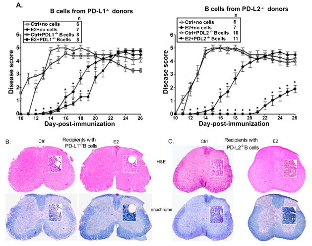
Figure 6. PD-L1 but not PD-L2 on B cells is critical for E2-mediated protection against EAE.
Female PDL1−/− and PD-L2−/− mice that served as donors of B cells were immunized with mouse (m)MOG35-55 peptide in Complete Freund’s adjuvant (CFA) with no Ptx administration. Seven to 8 week old μMT−/− mice that served as recipients of B cells were either sham-treated (control) or implanted with E2 pellets one week prior to B cell transfer and immunization with MOG35-55 peptide in CFA/Ptx. Recipient mice were monitored for signs of clinical EAE. A) Mean disease scores from 2–3 independent experiments with 3–5 mice/group/experiment. *p ≤ 0.05, compared to the respective control mice (i.e. E2-implanted with no cells vs. controls with no B cells and E2-implanted recipients with B cells from the appropriate donors vs. control recipients with B cells) (Mann-Whitney U test). Histopathological evaluation of spinal cords from B) PD-L1−/−, C) PD-L2−/− B cell-recipient μMT−/− (control and E2-implanted) mice (day 26 post-immunization). Spinal cords from each group, collected on day 26 post-immunization, were fixed in PFA and embedded in paraffin. 10 μm thick transverse sections from different regions of the spinal cord from each of the groups were stained with Hematoxylin & Eosin to enumerate infiltrating leukocytes and with Eriochrome cyanine to visualize the extent of demyelination (magnification 50 times and 200 times (inset)). Sections are representative of 2 experiments (n=3–5/group/experiment).

