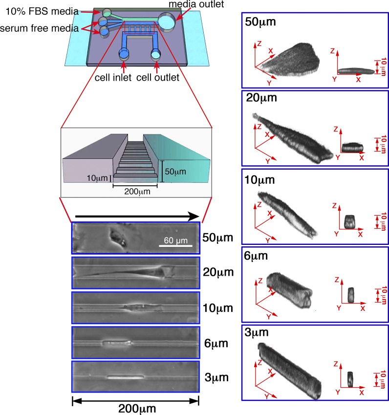
Figure 1.
Overview of the microchannel device. Schematic of the migration chamber bonded to coverslips (light blue), with inlet ports for serum-free media or cells (dark blue) or FBS (10%; green). Also shown is a close-up detailing the dimensions of the microchannel array, along with phase contrast and 3D reconstruction images of CHO-α4WT cells in microchannels of different widths. The black arrow (above the phase images) indicates the direction of migration.

