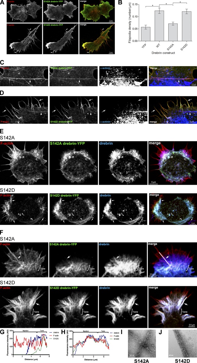
Figure 4.
Phosphorylation of drebrin at S142 is necessary and sufficient to relieve the intramolecular repression of F-actin bundling. (A) Immunofluorescence confocal images of COS-7 cells transfected with cDNA encoding YFP-tagged drebrin (green) mutated at S142 changing S to A (S142A) or S to D (S142D). Cells were labeled with phalloidin for F-actin (red). S142A drebrin-YFP only weakly induces F-actin containing filopodia, whereas S142D drebrin-YFP strongly induces filopodia (arrows). (B) Quantification of the number of filopodia per unit length of cell perimeter (filopodia density) in COS-7 cells transfected with cDNA encoding YFP, YFP-tagged drebrin (WT), and YFP-tagged drebrin mutated at S142 changing S to A (S142A) or S to D (S142D). Values are mean ± SEM (error bars) of 10 or more cells per transfection from three independent experiments. Significant differences (unpaired Student’s t test): *, P < 0.0001. (C and D) Immunofluorescence confocal images of COS-7 cells transfected with cDNA encoding S142A drebrin-YFP (C, green) or S142D drebrin-YFP (D, green) and labeled with phalloidin for F-actin (red) and antibody against α-actinin to label stress fibers (blue). S142A drebrin-YFP localizes to stress fibers (arrowheads) while S142D drebrin-YFP localizes to filopodia (arrows) and not to stress fibers (arrowheads). (E and F) Immunofluorescence confocal images of stage I cortical neurons (E) or growth cones from stage II/III cortical neurons (F) transfected with cDNA encoding S142A drebrin-YFP or S142D drebrin-YFP. Neurons were labeled with phalloidin for F-actin (red) and antibodies against GFP to visualize YFP-tagged drebrin (green) and drebrin (blue). S142A drebrin-YFP targets F-actin puncta (arrowheads) and actin arcs (curved arrows) in stage I neurons (E) and the Tzone in growth cones (F), whereas S142D drebrin-YFP localizes to F-actin puncta (arrowheads), filopodia (arrows), and actin arcs (curved arrows) in stage I neurons (E) and filopodia (arrows) in growth cones (F). (G and H) Fluorescence intensity in arbitrary units (AU) of F-actin (red), S142A drebrin-YFP (G, green), or S142D drebrin-YFP (H, green) and drebrin (blue) along the corresponding line plots shown in F (merge). S142A drebrin-YFP is mainly localized to the Tzone, whereas S142D drebrin-YFP is mainly localized to the base of the filopodium. Note that the drebrin level in the base of the filopodium is similar to that in the Tzone. (I and J) Electron micrographs of negatively stained F-actin from in vitro F-actin cosedimentation assays after addition of drebrin S142 mutant constructs. (I) After addition of S142A drebrin, thin, loose bundles of F-actin are visible, similar to those seen with wild-type drebrin (see Fig. 2 E). (J) After addition of S142D drebrin, thin, tight bundles of F-actin are visible. Bars: (A, D, and E) 5 µm; (C and F) 2.5 µm; (I) 100 nm; (J) 40 nm.

