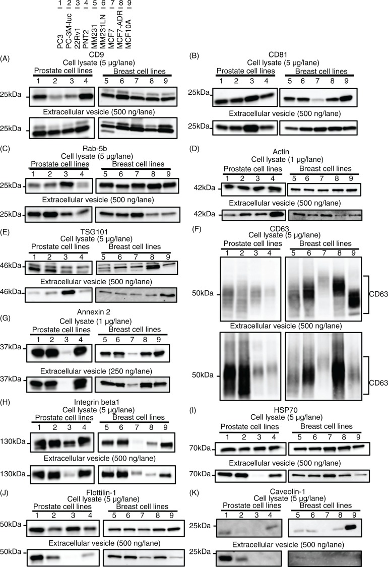
Fig. 4.
Immunoblotting analysis for conventional markers of EVs derived from prostate cell lines and breast cell lines. Proteins from whole cell lysates (upper panel) or EVs (lower panel) were separated on SDS-PAGE gels, followed by Western blotting using antibodies against 11 different EV markers. A 1 µg sample of cell lysate was used for the detection of Annexin 2 and actin. The other markers were detected using 5 µg of whole cell lysate. A 250 ng sample of EV proteins was used for the detection of Annexin 2. The other markers were detected using 500 ng of EV proteins. CD63, CD9, CD81 and Integrin beta 1 were detected under non-reducing conditions. Lane 1: PC3 cells; lane 2: PC-3M-luc cells; lane 3: 22Rv1 cells; lane 4: PNT2 cells; lane 5: MDA-MB-231-luc-D3H1 cells (MM231); lane 6: MDA-MB-231-luc-D3H2LN cells (MM231LN); lane 7: MCF7 cells; lane 8: MCF7-ADR cells and lane 9: MCF10A cells.

