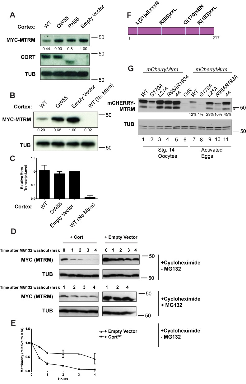
Figure 3. Cort expression leads to proteasome-mediated degradation of Mtrm in cell culture.
(A) Western blots showing levels of Mtrm and Cort in transfected Kc167 cells. pMT-cort and pMT-6×myc-mtrm were transfected into Kc167 cells. The form of transfected Cort is indicated above each lane. Only wild-type Cort leads to decreased levels of tagged Mtrm protein. The RH65 mutation results in a premature stop codon in Cort. Myc-Mtrm band intensity is quantified below the Myc-Mtrm panel. Band intensity is normalized to tubulin and is expressed relative to empty vector. (B and C) Cells transfected with pMT-6×myc-mtrm (lanes 1–3; lane 4 transfected with pMT-empty in place of mtrm) and the indicated form of Cort (WT, QW55, or pMT-empty) were split and subjected to both Western blot (B) and quantitative PCR (C). Myc-Mtrm band intensity is quantified as in (A). For qPCR, mtrm transcript levels are normalized to actin5c and shown relative to empty vector. (D) Western blot showing Mtrm protein levels over time. Time indicates hours post-MG132 washout. Rate of Matrimony degradation is faster in the presence of Cortex versus empty vector. The rate of degradation is slowed in continued presence of MG132. (E) Quantification of –MG132 blot in (D). The 1-, 2-, and 4-h time points are averages of two experiments. Mtrm amount is normalized to tubulin and shown relative to amount at the 0 h time point. (F) Illustration of candidate APC/C recognition motifs. (G) The L21A mutation stabilizes mCherry-Mtrm in embryos. Western blots of stage 14 oocytes and fertilized eggs (1 h collection) are shown. The 4A mutant consists of L21A, R95A, R193A, and H94A (a mutation in a possible APC/C initiation motif [54]). Percentage below mCherry activated egg lanes indicates remaining protein left, normalized to tubulin, and relative to amount at stage 14. Asterisk denotes cleavage product due to hydrolysis of acylimine linkage in the mCherry tag [55]. Myc-Mtrm was detected using anti-Myc antibody (A, B, D) and mCherry-Mtrm was detected using anti-RFP (G).

