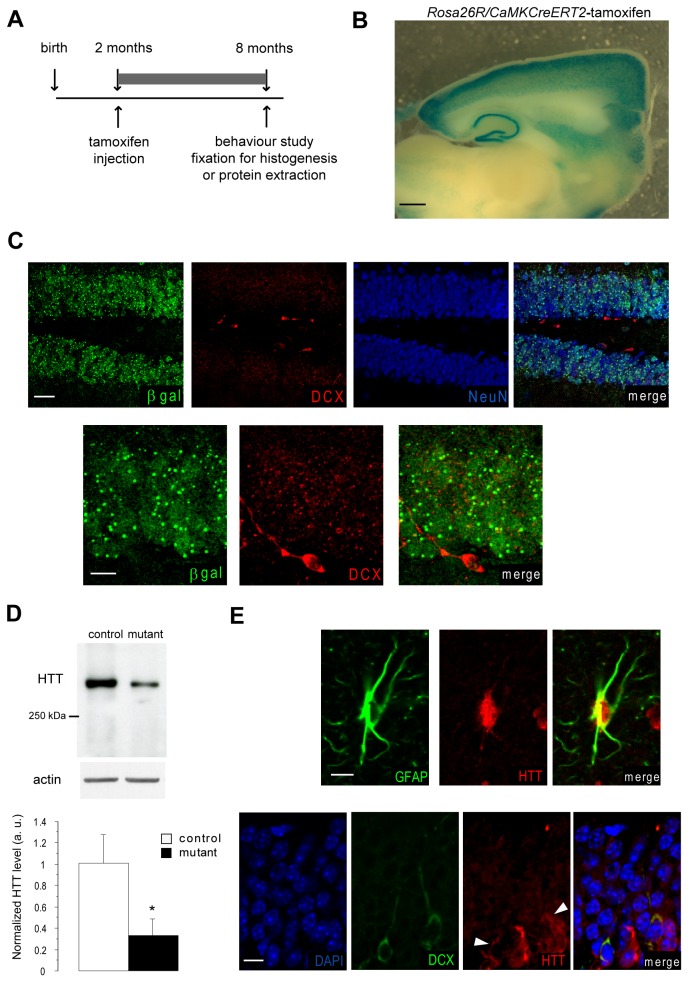
Figure 1. Tamoxifen injection in CaMKCreER T2 mice activates Cre expression in mature but not newborn hippocampal neurons.
(A) Design of experiments performed on CaMKCreER T2 ;ROSA26R, CaMKCreER T2 ; Hdh flox/flox and WT; Hdh flox/flox mice. (B) Whole mount X-gal staining of a parasagittal brain section of a CaMKCreER T2 ;ROSA26R mouse injected with tamoxifen. Scale bar: 500 µm. (C) Immunofluorescence in the hippocampal region of tamoxifen-injected CaMKCreER T2 ;ROSA26R mouse with antibodies recognizing β-galactosidase, DCX and NeuN. Scale bar for upper panel: 30 µm ; scale bar for lower panel: 10 µm. (D) Representative Western-blot of hippocampal proteins extracted from mutant and control mice 6 months after tamoxifen injection and incubated with anti-Htt (4C8) and anti-actin antibodies. Data are the mean +/- SEM of the ratios Htt/actin, normalized so that the mean value of controls is equal to 1 (n= 4-5 per group). * p<0.05 (E) Hippocampi of mutant mice 6 months after tamoxifen injection were co-immunostained with antibodies recognizing GFAP or DCX and HTT (4C8). White arrows indicate HTT expression in DCX positive neurons. Scale bar for upper panel : 10 µm; scale bar for lower panel : 8 µm.

