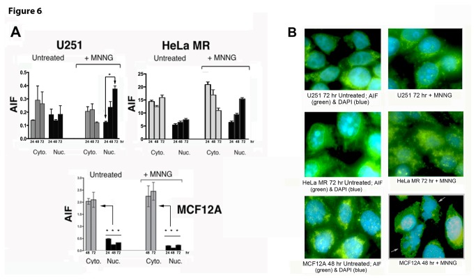
Figure 6. Apoptosis Inducing Factor (AIF) translocates to the nucleus in cancer cells (U251 and HeLa MR) but not in normal cells (MCF12A) after MNNG exposure.
U251 and HeLa MR tumor cells exhibit increased AIF traversal into the nucleus 48-72 hr after MNNG (0.2 µM). MCF12A normal human cells do not exhibit increased traversal of AIF into the nucleus up to 96 hr after equitoxic MNNG (8 µM). A. AIF detected by cell fractionation and quantitative immunoblotting. Each fluorescent band was measured against a loading control (cytoplasmic GAPDH or nuclear lamin) in the same lane. Asterisks (*) denote statistically significant differences at P < 0.05 between the amount of AIF measured within nuclear extract at 72 hr and at 24 hr after MNNG treatment (U251 + MNNG), or between each nuclear extract and cytoplasmic extract concentration at 72 hr (MCF12A). Histograms produced by Prism GraphPad software, error bars indicate SD. Statistical significance determined by student t-test using Prism GraphPad software. Each experiment performed a minimum of two times. B. Microscopic immunofluorescence (100X) using AIF antibody (green) and DAPI (blue). HeLa MR and U251 cells exhibit maximum AIF within the nucleus at 72 hr. MCF12A cells do not exhibit AIF traversal to the nucleus, but do exhibit apoptotic nuclei at 48 hr as indicated by the two white arrows in the lower left photomicrograph.

