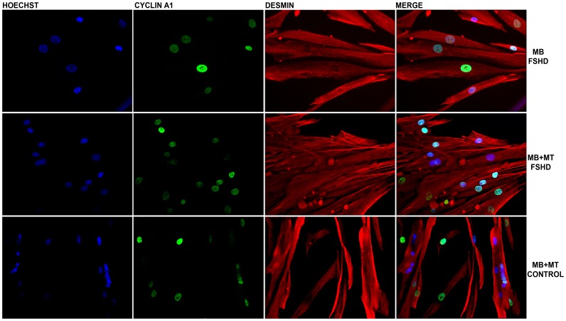
Figure 3. Immunostaining of myoblasts (MB), and myoblasts and myotubes (MB-MT).
(40×magnification) from one FSHD patient and one healthy control. Desmin (red), cyclin A1 (green), Hoechst (blue) staining. Cyclin A1 was detected in nuclei of myoblasts and myotubes derived from both FSHD patients and healthy controls.

