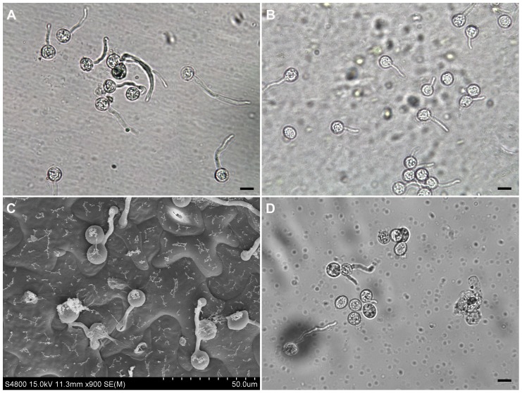
Figure 1. The Phytophthora capsici cysts germinating on different surfaces.
The cysts were germinated on a cellophane membrane that was placed on the top of a Nicotiana benthamiana leaf (A), or directly on N. benthamiana leaves (B-C), or directly in water (D). Photos were taken at 70 (A), 60 (B-C) and 90 (D) min post-inoculation. To make samples for observation in (B), free water around the inoculation sites was absorbed away by filter paper and germinating cysts with some leaf tissue were peeled off using the sticky tape method [79]. The cysts germinating directly on N. benthamiana leaves were also observed using Cryoscanning electron microscopy (Hitachi S-4800 SEM) according to the instruction manual (C). The germinating cysts (A, B and D) were observed under an Olympus microscope. Scale bar = 10 µm.

