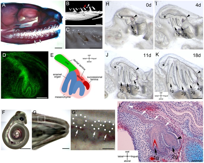
Figure 1. Slice organ culture of snake mandibles allows the formation of several generations of teeth.
(A–C) Newborn corn snake (Pantherophis guttatus). (A) Alcian blue-Alizarin red skeletal staining of the head. (B) Mandible microCT scan. (C) Oral view of erupted teeth. (A–B) White arrows indicate functional teeth. (B) Red arrow and arrowhead point to replacement teeth. The arrowhead indicates the younger replacement tooth. (D) 3-day cultured corn snake dental organ. Fibrillar-Actin (green) is used to show the outline of the developing tooth germs. (E) Schematic of the regions of the dental organ represented in D. (F) 30-days post-oviposition snake embryo. (G) Mandible from embryo in C. (G′) Magnification of the framed area in G, dental organs can be identified throughout the mucosa indicated by arrows and dotted circles in the inset. (H–K) 18-day culture period. Several generations of teeth develop over this period. (L) Histology of dental lamina after 10 days in culture. (H–L) Black arrows: tooth germs; 1 g, 2 g, 3 g, 4 g, are 1st, 2nd, 3rd, and 4th generations of teeth. Black arrowheads: successional lamina. White arrows: dental lamina. White arrowhead: origin of dental lamina from the oral epithelium. Scale bars: (A) 100 µm, (C,F) 1 cm, (G) 1 mm, (D,E) 500 µm, (G′,H–K) 250 µm, (L) 100 µm.

