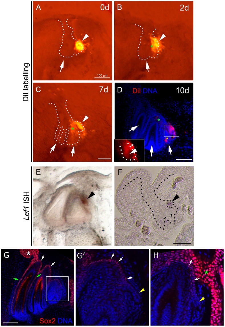
Figure 2. Fate and expression pattern of the dental lamina in the snake.
(A–D) 10-day culture of the snake dental organ labelled with DiI. (A, B, C) fluorescence microscopy and (D) confocal optical section. (A) 0-day culture. DiI labelling was performed at the region of the successional lamina (arrowhead). (B) 2-day culture. (C) 7-day culture. (B,C) The label remains in the region of the successional lamina (arrowhead) and in the forming second tooth (green asterisk). (D) Day 10. Magnification in D shows the framed area. Label is present in the second (green asterisk) and third (yellow asterisk) generation tooth germs and is retained in the successional lamina (arrowhead) and adjacent mesenchyme (white asterisk). (E–F) Pantherophis guttatus Lef1 mRNA is located in the successional lamina region (arrowhead), as observed by whole mount (E) and section (F) in situ hybridization. (G,G′,H) Sox2+ cells are found in the oral (asterisk) and aboral dental lamina (white arrows) and outer enamel epithelium (grey arrows), but are excluded from the successional lamina (yellow arrowhead in G′, H). (G′) magnification of the region indicated in G. (H) Lingual plane showing the connection of the dental lamina with the oral epithelium. Scale bars: 100 µm.

