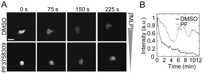
Figure 5. PAK inhibition alters neutrophil calcium signaling.
Human neutrophils were loaded with the intracellular Ca2+ dye fluo-4 (2 µM) and plated on fibronectin-coated surfaces. After treatment with DMSO or PF3758309 (10 µM), neutrophil chemotaxis was induced by the addition of fMLP in an Insall chamber. Intracellular Ca2+ release was monitored every 5 s for 15 min by fluorescent microscopy. (A) Time-lapse images after the peak Ca2+ spike were shown. (B) Ca2+ spikes in cells treated with 0.1% DMSO (black line) or PF3758309 (PF; PAK inhibitor, dotted line) were quantified and presented as mean intensity of at least 5 neutrophils in a field of view. Data shown is representative from 3 independent experiments. Scale ba = 20 µm.

