
Figure 5. Immunohistochemical analysis of the stomach of Pipistrellus pipistrellus.
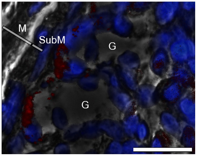
Picture overlay of labeling of the α-AMCase antibody (red); DAPI counterstained (blue) and phase contrast demonstrating positive labeling of the antibody at the bottom of the gastric glands. Bar = 50 µm. G = gastric gland, M = mucosa, SubM = submucosa.