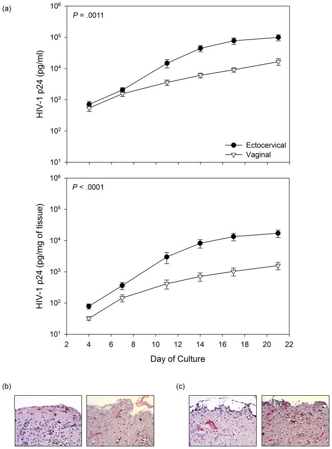
Fig. 1.
HIV-1 replication and infection of fresh ectocervical and vaginal tissue. Paired ectocervical and vaginal tissue was collected from 42 participants. The tissue was exposed to HIV-1, wash, and cultured for 21 days. Supernatant was collected and replenished every 3 to 4 days. (a) HIV-1 replication was monitored by p24gag ELISA in the culture supernatant. The p24 values were summed over day 4 through 21 and presented as the mean ± SD of 41 participants (one participant’s tissue was contaminated). The p24 values were from non-weight adjusted (upper panel) or weight adjusted (lower panel) p24 values. Embedded probability values for the paired comparison of cumulative p24 at day 21. Representative immunohistochemistry staining for p24 infected cells from cervical (b) and vaginal (c) tissue collected at study end is shown.

