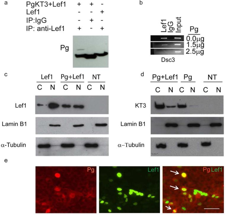
Figure 3. Pg co-localizes in the nucleus with Lef1 and disrupts TCF/Lef transcription factor binding to the Dsc3 promoter.
(a) Co-Immunoprecipitation (Co-IP) assays demonstrating an interaction between Pg and Lef-1. (b) ChIP assays demonstrating that increasing amounts of Pg (measured in g plasmid transfected) interfere with the binding of Lef-1 to the Dsc3 promoter. Input, chromatin used for immunoprecipitation; IgG, IP with unspecific IgG. (c, d) Western blot analysis of MDCK cells transfected with Pg and Lef-1 (transfection constructs shown on top; NT, not transfected). The nuclear (N) and cytoplasmic (C) distribution of the proteins is shown. Antibodies used to detect Lef-1 and the KT3-tagged plakoglobin construct are shown on the left sides of the blots. Our Lef-1 antibody does not detect endogenous Lef-1 expression in MDCK cells. Lamin B1 (nuclear fraction) and α-tubulin (cytoplasmic fraction) antibodies were used as controls. (e) Immunofluorescence microscopy of MDCK cells transfected with Pg and Lef-1. The antibodies used for staining are indicated. Note the nuclear co-localization of Pg and Lef-1 in several cells (arrows). Bar, 50 μm

