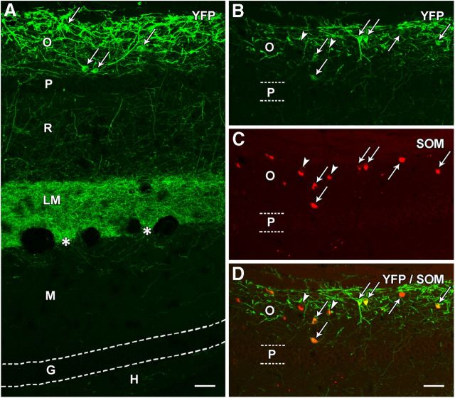
Figure 4.
Localization of eYFP in a normal SOM-Cre mouse following viral delivery of Cre-dependent ChR2-eYFP construct to s. oriens. A, At 3 weeks after injection, eYFP labeling is prominent in neuronal cell bodies (examples at arrows) and processes in s. oriens (O) of CA1. A few labeled processes extend through the adjacent pyramidal cell layer (P) and traverse s. radiatum (R). A dense plexus of fibers is present throughout s. lacunosum-moleculare (LM) but ends sharply at the hippocampal fissure (asterisks). Very limited fiber labeling is evident in the adjacent molecular layer (M) of the dentate gyrus. Virtually no labeling is present in the granule cell layer (G) or hilus (H) of the dentate gyrus, indicating that the viral vector did not reach the hilar region where additional Cre-expressing neurons are located in normal mice. B–D, In sections immunolabeled for SOM, essentially all eYFP-expressing neurons were SOM neurons (arrows), as demonstrated by the merged images. However, a few SOM neurons in the region did not appear to be labeled for eYFP (arrowheads). Scale bars: 50 μm.

